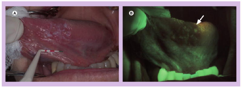
Figure 1. Wide-field autofluorescence imaging.

(A) A white light image of the ventral tongue of a patient with an oral premalignant lesion, which, when biopsied, was confirmed to be severe dysplasia and (B) a corresponding autofluorescence image obtained with the VELscope® (LED Dental Inc., BC, Canada). The arrow indicates the region of fluorescence visualization loss and biopsy location.
Reprinted from with permission from [26] © SPIE 2006.
