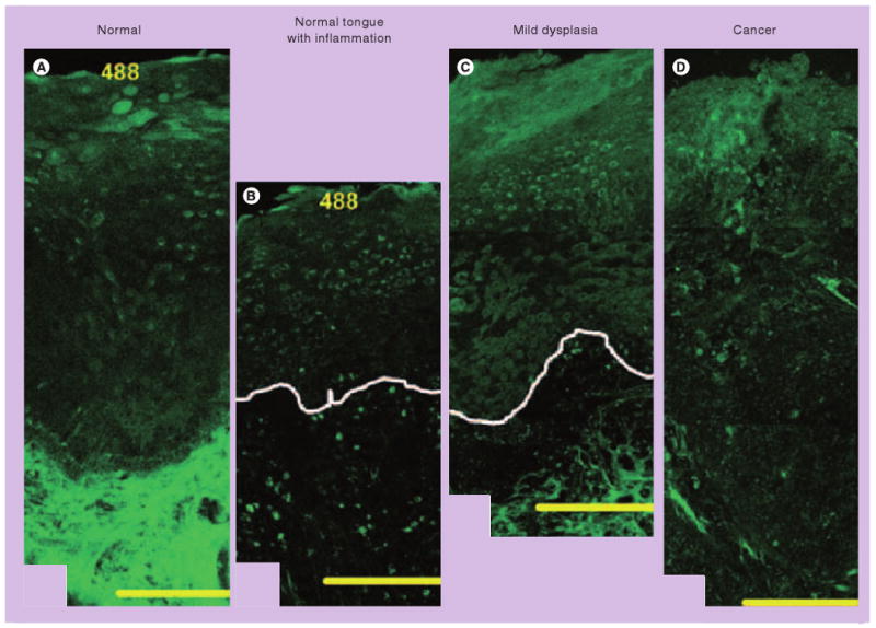
Figure 4. Confocal fluorescence images at 488 nm excitation of fresh organ cultures.

(A) The origins of epithelial and stromal fluorescence in the normal tongue. Adjacent images of lesions in the tongue illustrate the changes in epithelial and stromal fluorescence associated with severe inflammation (B), and mild dysplasia and mild-to-moderate inflammation (C). White lines indicate the approximate location of the basement membrane. (D) A fluorescence image of poorly differentiated carcinoma in the palate. Scale bars: 200 μm. Inflammatory lesions show decreased epithelial fluorescence, whereas dysplastic lesions display increased epithelial fluorescence compared with normal oral tissue. Stromal fluorescence in both inflammatory and dysplastic lesions drops significantly. Thus, wide-field fluorescence imaging, a technique that captures autofluorescence generated primarily in the stroma, may fail to distinguish inflammation from precancerous lesions. One approach to improve diagnostic accuracy is to obtain images from more superficial layers using high-resolution imaging systems.
Reprinted with permission from [52].
