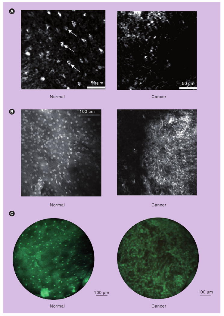
Figure 5. High-resolution imaging.

(A) High-resolution images obtained in vivo with a fiber optic confocal reflectance microscope. Small, regularly spaced epithelial cell nuclei are clearly visible in the intermediate squamous epithelium of the normal site (left). A confocal image of oral squamous cell carcinoma is characterized by disordered tissue structure (right). Scale bars: 50 μm. (B) High-resolution fluorescence images after topical application of acriflavine hydrochloride in ex vivo specimens. Normal mucosa with regular configuration of cell nuclei (left) and in an invasive carcinoma of the floor of the mouth showing different sizes of nuclei (right) (imaging plane depth: ∼50 μm). Scale bar: 100 μm. (C) High-resolution fluorescence images of oral tissue after topical application of proflavine obtained with a fiber optic fluorescence microendoscope, demonstrating qualitative differences between normal and cancerous tissue. Scale bars: 100 μm. The high-resolution microendoscope used to obtain images in (C) is portable, battery-powered and has been used in vivo in a variety of clinical settings.
(A) Reprinted with permission from [57] © Elsevier (2008). (B) Reprinted with kind permission from [59] © Springer Science+Business Media (2009). (C) is reprinted with permission from [60] © Optical Society of America (2007).
