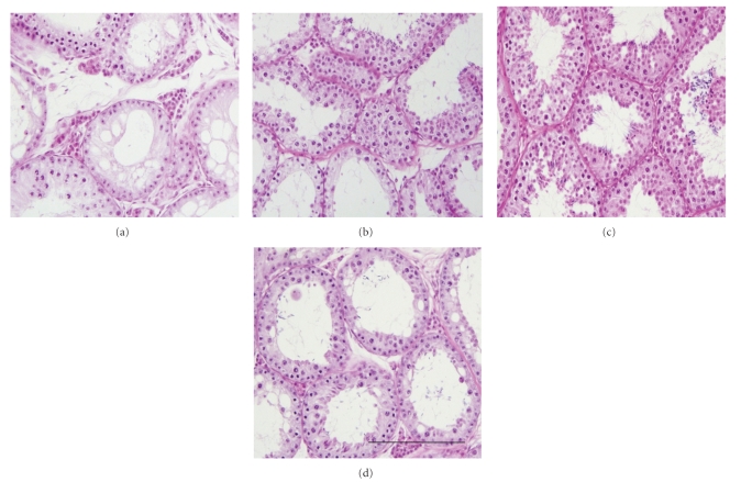
Figure 1.
Histological photomicrographs of testis tissue xenografts recovered from recipient mice at 8 months post grafting. Representative xenografts from the group of recipient mice receiving 2 (a), 4 (b), 8 (c), or 16 (d) testis tissue fragments. The grafts from mice receiving 16 fragments were overall larger and more developed than those from mice receiving 2 fragments at the time of grafting. Scale bar = 200 μm.

