
Figure 1. The placenta is a hematopoietic organ that supports generation and proliferation of hematopoietic stem cells (HSC).
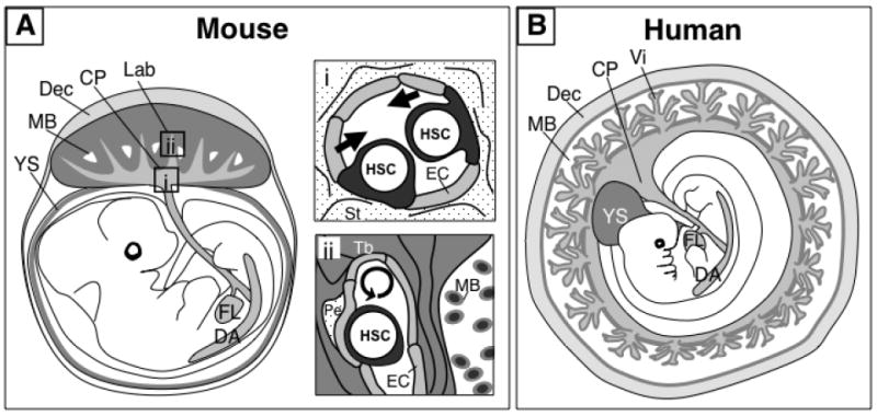
A) The mouse placenta consists of two distinct vascular regions that function as putative niches for HSC development. The chorionic plate (CP) harbors large fetal blood vessels that are contiguous with the umbilical cord vessels and connect with the fetus. These blood vessels are potential sites of HSC generation (i). The labyrinth (Lab) region contains an extensive network of small fetal blood vessels that are surrounded by trophoblasts (Tb) lining the maternal blood spaces (MB). The labyrinth is the site of fetal-maternal exchange, and may serve as a niche for HSC expansion (ii). B) The human placenta contains also two vascularized regions, the chorionic plate and the villi (Vi). The villi are physiologically analogous with the labyrinth of the mouse placenta and contain hematopoietic stem/progenitor cells in the vascular and perivascular locations. It is hypothesized that the large vessels of the chorionic plate in the human placenta also have de novo hematopoietic potential. Although the macroscopic organization of the placenta and other extra-embryonic tissues differ between humans and mice, it is likely that similar cellular and molecular mechanisms coordinate placental hematopoiesis in both species. Dorsal aorta (DA), yolk sac (YS), fetal liver (FL), decidua (Dec), endothelial cell (EC), stroma (St), pericyte (Pe).