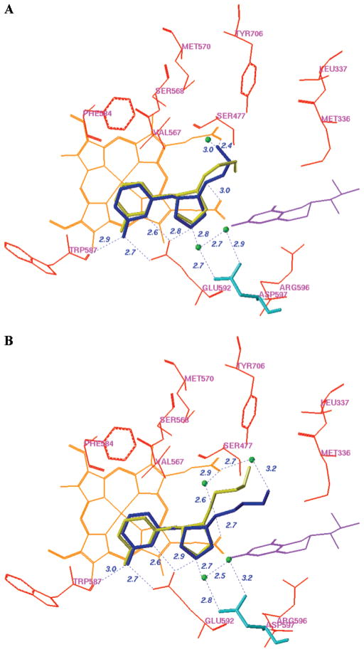
Figure 7.
Superimposition of the binding conformation (blue) and predicted bioactive conformation (yellow) of 4 (A) and 6 (B) in the active site of rat nNOS. The heme (orange), H4B (violet), and structural water (green) involved in the binding of 4 and 6 to nNOS are shown. The distances of some important H-bonds between the residues, structural water, cofactors, and inhibitors are given in angstroms (Å).

