
Figure 1. Crystal structures of members of the four recognised AB5 toxin families.
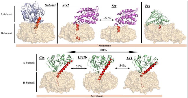
The B-subunit is represented as molecular surface. The A-subunits of SubAB, Ctx and LT, Stx and Ptx are shown in cartoon representation and coloured according to the respective catalytic activity (Light blue for subtilase activity, light green for ADP-ribosylase activity and purple for RNA N-glycosidase activity). The common structural element (Helix A2) is coloured in red, and the level of sequence identity of the A-subunit inside a family is also indicated.