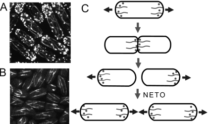
Figure 1. Images of yeast cells (Jian-Qiu Wu, Ohio State University) and yeast growth pattern.
(A) Images of the actin cytoskeleton in cells expressing GFP-CHD which binds to the sides of actin filaments. Actin cables and actin patches are seen distributed in monopolar and bipolar patterns. (B) In cells expressing GFP-atb2, microtubule bundles run across the cell. (C) Cartoon showing the redistribution of the actin cytoskeleton during the cell cycle. Prior to cytokinesis, actin accumulates at growing tips; during mitosis it accumulates in the middle; daughter cells start to grow in a monopolar manner and transition to bipolar growth at new end take-off.

