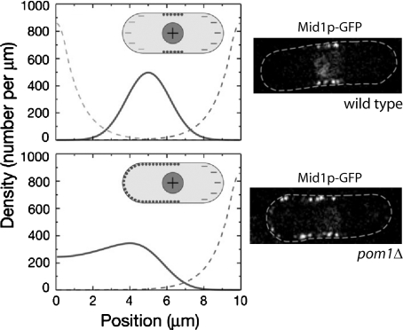
Figure 5. Results of model of positioning of Mid1p in nodes in the middle of the cell and experimental images (Padte et al., 2006).
The nodes are precursor components of the contractile ring. The model involves activation of Mid1p in the nucleus, deactivation near cell tips (dashed line), and binding of active Mid1p to the membrane. Top graph shows simulated distribution of membrane-bound Mid1p (solid line) as a function of position along the cell. Bottom graph shows membrane-bound Mid1p in cells lacking one of the polar inhibitors, Pom1p. Micrographs show the corresponding experimental images. Images reproduced with permission.

