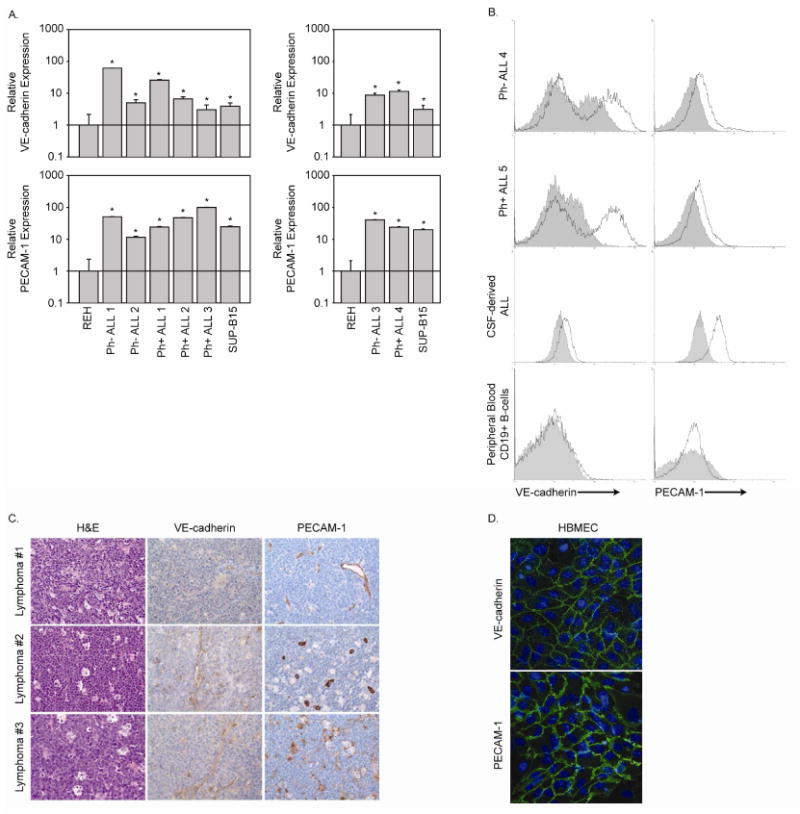
Figure 3. Primary ALL expresses cell surface VE-cadherin and PECAM-1.

A. RNA from leukemic cells isolated from leukaphoresis (Ph- ALL1, Ph- ALL 2, Ph- ALL 3, and Ph+ ALL 1) and bone marrow aspirates (Ph+ ALL 2, Ph+ ALL 3, and Ph+ ALL 4) was examined for VE-cadherin and PECAM-1 expression by real-time RT-PCR. RNA from REH and SUP-B15 cells was included as negative and positive controls, respectively. Data are expressed as fold increase over the gene expression levels in REH cells. B. Leukemic cells derived from bone marrow aspirates (Ph- ALL 4 and Ph+ ALL 5) and cerebrospinal fluid (CSF) as well as peripheral blood CD19+ B-cells from a healthy donor were evaluated for cell surface VE-cadherin and PECAM-1 expression by immunostaining and flow cytometric analysis (solid line represents specific primary antibody while shaded histograms represents the isotype matched control). C. High-grade B-cell lymphoma biopsy samples were evaluated for VE-cadherin and PECAM-1 expression by immunohistochemistry. Samples were also stained with hematoxylin and eosin. Photomicrographs were taken at 400× magnification. D. HBMEnd grown to confluence on glass coverslips were fixed and immunostained to detect the adherens junction proteins VE-cadherin and PECAM-1. Cell nuclei were stained using DAPI.
