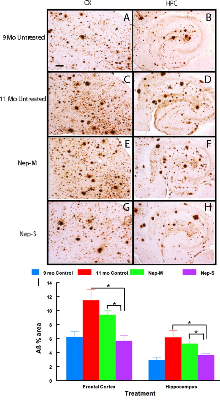
Figure 7.

Immunohistochemical Aβ deposition is blocked in APP+PS1 mice infused with GFP monocytes transfected with NEP-s but not NEP-m. Brain sections harvested 1 d after the last infusion were stained with antibody against Aβ to estimate the amount of diffuse and compacted Aβ deposition. Representative micrographs of staining from frontal cortex (A, C, E, G) or hippocampus (B, D, F, H) are shown for each of the four treatment conditions (indicated to the left of each pair of micrographs). I shows quantification of percentage area of Aβ immunostaining in the frontal cortex and hippocampus. Sample size per group = 7–9. Statistical analysis was performed using one-way ANOVA with Fisher's LSD multiple-comparison test. Brackets between bars signify statistical significance (p < 0.05). Values are mean ± SEM (standard error of mean). Magnification, 40×. Scale bar, 120 μm for all panels.
