
Figure 5. Reduced α- and γ- sarcoglycan expression in immature biglycan null mice.
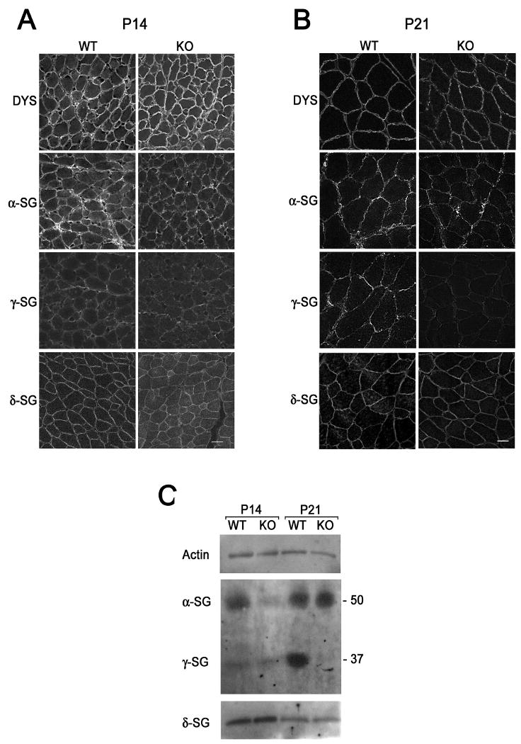
Immunohistochemical analysis of P14, A., and P21, B., mouse muscle. Sections of quadriceps femoris from congenic P14 wild type and biglycan null (KO) mice were sectioned, mounted on the same slides and immunolabelled for dystrophin or α-, γ-, or δ- sarcoglycan. A. α-Sarcoglycan levels at the sarcolemma of P14 biglycan null muscle are selectively reduced in the biglycan null as compared to wild type muscle. B. γ-Sarcoglycan expression is reduced in P21 biglycan null mice, while α-sarcoglycan is unchanged. The level of dystrophin and δ-sarcoglycan is the same at both ages. Scale bars = 10 μm. C. Biochemical analysis of sarcoglycan expression. α- and γ- Sarcoglycan expression is reduced at distinct postnatal ages in skeletal muscle membranes from immature mice. KCl-washed skeletal muscle membranes (3 μg) from P14 or P21 congenic wild type or biglycan null mice (KO) were separated via SDS-PAGE. Western blotting was performed for α-, γ-, or δ- sarcoglycan or actin (loading control). Western blotting for α- and γ- sarcoglycan was performed on the same gel. Western blotting for δ-sarcoglycan and actin was performed in parallel on the same gel. α- Sarcoglycan levels are reduced in P14 biglycan null membranes while γ-sarcoglycan levels are reduced in P21 biglycan null membranes. Equivalent levels of δ-sarcoglycan and actin expression are seen at both ages. Similar results were observed in muscles from two other sets of mice.