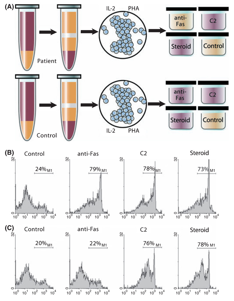
Fig 2.
ALPS apoptosis assay. Peripheral blood mononuclear cells are isolated from a patient, and T cells are activated with mitogen (phytohae-magglutinin, PHA) and expanded with IL-2 in culture for approximately 28 d (Fisher et al, 1995). In normal individuals, mitogen activation and expansion results in upregulation and priming of the Fas apoptotic pathway. When normal T cells are exposed to anti-Fas IgM monoclonal antibody in vitro, they undergo rapid cell death and apoptosis (Teachey et al, 2005). Because patients with ALPS have a defect in this pathway, the cells do not die after exposure to anti-Fas IgM monoclonal antibody (Sneller et al, 2003). Dexamethasone and ceramide (C2) are used as positive controls. (A) Red wells depict cell death after treatment with anti-fas IgM, C2, or dexamethasone. Yellow wells depict lack of cell death with similar treatment. (B) Normal control demonstrating cell death with anti-Fas IgM, ceramide, and dexamethasone as depicted by histogram for 7-Aminoactinomycin D. (C) Prototypical patient with ALPS demonstrating cell death with exposure to ceramide and dexamethasone, but no increased death over baseline with exposure to anti-Fas monoclonal antibody. Data collected under Institutional Review Board approved protocol. © Sue Seif, MA (used with permission).

