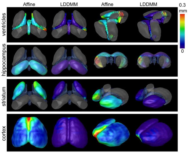
Figure 3.
Accuracy of affine and nonlinear registration on the surfaces of ventricles, hippocampus, and striatum by using manual segmentation as standard. In vivo MRI images from six 12-week-old R6/2 mouse brains were normalized to our atlas image by using affine and nonlinear transformation (Miller et al., 2002). The segmentation in the atlas image was transformed into each subject image by affine or LDDMM transformations. The distances between the structural boundaries reconstructed from automated segmentation and surfaces reconstructed from manual segmentation results are visualized on the structural surfaces.

