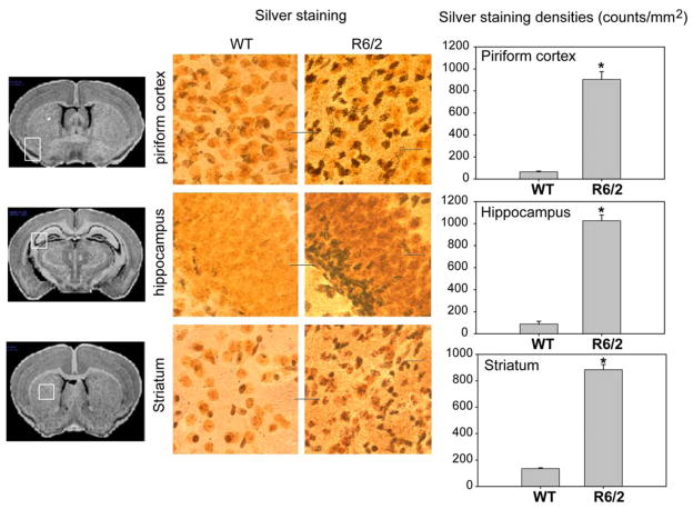
Figure 7.
Neurodegeneration was evident in R6/2 mice by silver staining. Silver staining of brains of 12-week-old control mice (WT) and R6/2 mice. Note that silver-positive dark bodies represent cells undergoing neurodegeneration. The degenerating neurons were detected in hippocampus, striatum and piriform cortex (middle column), which regions showed atrophy in MRI measures (right column). Scale bars=20 μm. Quantitative silver staining densities in different brain region as indicated in piriform cortex (left panel), hippocampus (middle panel) and striatum (right panel). *p<0.01 compared to the values of those in nontransgenic control wild type (WT) mice. One-way ANOVA with posthoc tests were used.

