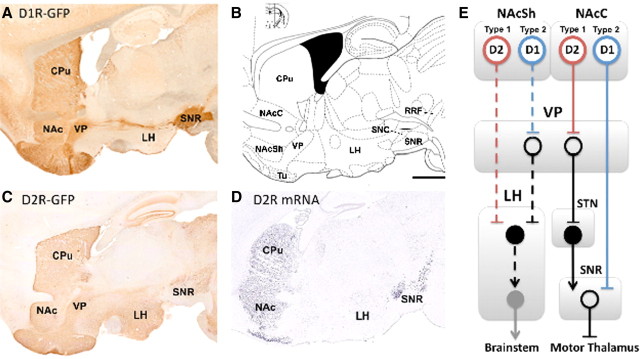
Figure 1.
A, C, Projections of D1-containing (A) and D2-containing (C) neurons in mouse brain, courtesy of GENSAT. B, Anatomical locations at interaural 1.08 mm defined by Paxinos and Franklin (2001). Bar = 1 mm. D, In situ hybridization for D2 mRNA, courtesy of the Allen Mouse Brain Atlas (Allen Institute for Brain Research, 2009). E, Simplified schematic of the known (solid lines) and hypothesized (dashed lines) output pathways of the nucleus accumbens. We propose that the type 1 neurons defined by Krause et al. (2010) are D2 MSNs and the type 2 neurons are D1 MSNs. Open circles, inhibitory neurons; filled circles, excitatory neurons. CPu, Caudate–putamen; NAc (Sh/C), nucleus accumbens (shell/core); Tu, olfactory tubercle; SN (R/C), substantia nigra (reticulata/compacta); RRF, retrorubral field; STN, subthalamic nucleus.

