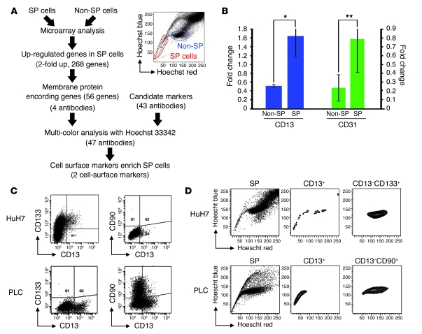
Figure 1. CD13 is a candidate marker of the SP fraction.
(A) The strategy used to identify cell-surface markers closely related to the SP fraction. We determined CD13 and CD31 as candidate markers for identifying SP cells. (B) Both CD13 and CD31 expression in HuH7 cells were compared in SP and non-SP cells by semiquantitative RT-PCR. Data represent mean ± SD from independent experiments of fractions differentially sorted by flow cytometry. *P < 0.01; **P = 0.076 versus non-SP fractions. The cut-off lines were determined using isotype controls. (C) Expression of CD13, CD133, and CD90 in HuH7 (upper panels) and PLC/PRF/5 cells (lower panels). Horizontal and vertical axes denote expression intensity. (D) The SP fraction is recognized as a “beak” appearing beside the G1 phase fraction. The relationship between CD13+ and CD13– cells and the SP fraction was studied using multicolor flow cytometry.

