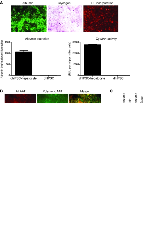
Figure 2. In vitro modeling of A1ATD using disease-specific human iPS cells.
(A) A1ATD disease-specific human iPS cells (dhIPSCs) differentiated to hepatocytes displayed functional activity characteristic of primary human hepatocytes, including the presence of intracellular albumin, glycogen storage (shown by PAS staining), LDL cholesterol uptake (shown by fluoresceinated LDL incorporation), albumin secretion, and active CytP450 metabolism. Error bars denote SEM. (B) Immunostaining analyses for expression of misfolded polymeric α1-antitrypsin using the polymer-specific 2C1 antibody (green) or an antibody that detects all forms of α1-antitrypsin (red) in A1ATD patient-specific and control human iPS cell–derived hepatocytes. Merged images are shown at right. (C) Endoglycosidase H (EndoH) digestion of A1ATD disease-specific human iPS cell–derived hepatocyte microsomal subcellular fraction, confirming retention of misfolded polymeric α1-antitrypsin protein within the endoplasmic reticulum. n = 3. (D) ELISA to assess the intracellular expression (cells) and secretion (medium) of all α1-antitrypsin and polymeric α1-antitrypsin protein in A1ATD patient-specific and control human iPS cell–derived hepatocytes after overnight proteasomal inhibition by MG132. Error bars denote SEM. n = 3. Original magnification, ×20 (A); ×40 (B).

