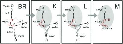
Figure 6.
Structural changes of the Asp-85–Thr-89 region on the basis of the present FTIR results. The distances represent those between two oxygen atoms in 1C3W (10). The shaded oval represents the cluster structure in the Asp-85–Thr-89 region. According to the present scheme, OD1 accepts the proton from the retinal Schiff base in M, whereas OD2 loses hydrogen bonds with water molecules.

