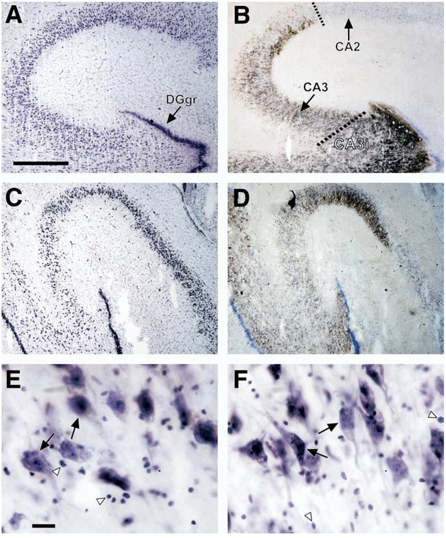
Figure 1.
Brightfield photomicrographs of coronal sections of the postmortem human hippocampal formation. (A) Cresyl violet–stained section from a 70-year-old male control subject (postmortem interval = 20 hours) and (B) an adjacent section processed by Timm staining. Note the intensely stained granule cell layer of the dentate gyrus (DGgr) in (A) and (B), and the clear demarcation in (B) between hippocampal subfields CA2 and CA3 afforded by the Timm staining. A dashed line identifies the border between CA2 and CA3, and the second dashed line shows the border between CA3 inserted within the dentate gyrus (CA3i) and CA3 external to the dentate gyrus. (C) Cresyl violet–stained section from a depressed 77-year-old man (postmortem interval = 26 hours) and (D) an adjacent section processed by Timm staining. Pyramidal neurons and glial nuclei of CA3 are highlighted (E, Control; F, MDD) with large black arrows and white arrowheads, respectively. The scale bars in (A) and (E) are 750 µm and 25 µm, respectively.

