Fig. 2.
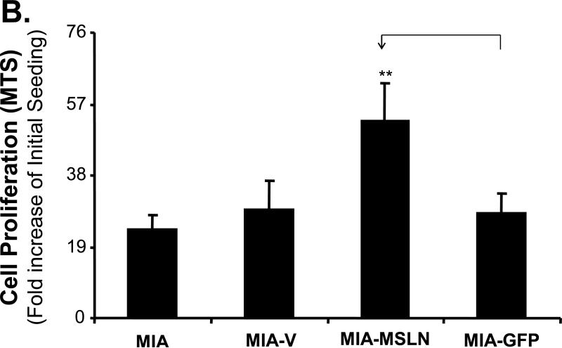
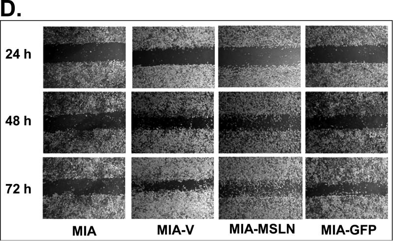
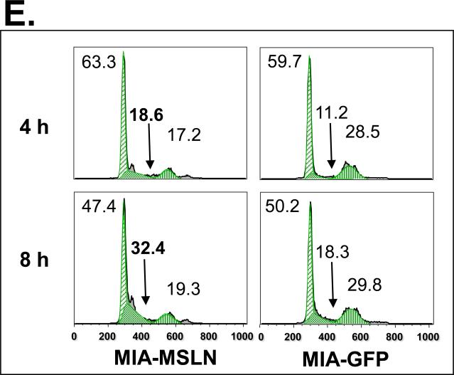
Effects of MSLN overexpression in MIA PaCa-2 cells on cell proliferation and migration. A. MSLN expression in stable cells by Western blot analysis. B. Cell proliferation by MTS assay. Cells were seeded in 96-well plates (2 × 10³ cells/well) and serum-starved for 24 h before changing to the growth medium with 2% FBS. Absorbance was recorded on day 6 after release from starvation at 490 nm. C. Cell migration by modified Boyden chamber assay. Stable cells were trypsinized and resuspended in growth medium (105 cells/500 μL) and added into the upper compartment of a migration chamber for 24 h. The cells were stained with Calcein-AM. The mean fluorescence reading after scraping of the cells at the top was divided by the reading before removal of the top cells, and the ratio was plotted. D. Cell migration by wound-healing assay. Confluent stable cells were scraped with a pipette tip and cultured in DMEM for 3 days. Pictures were taken daily. E. Cell-cycle analysis by flow cytometry. Confluent cells were serum-starved (0% FBS) for 24 h before changing to the growth medium with 2% FBS. Cells were collected at 4 h and 8 h, permeabilized, stained with propidium iodine (PI), and analyzed with flow cytometry. **p < 0.001.


