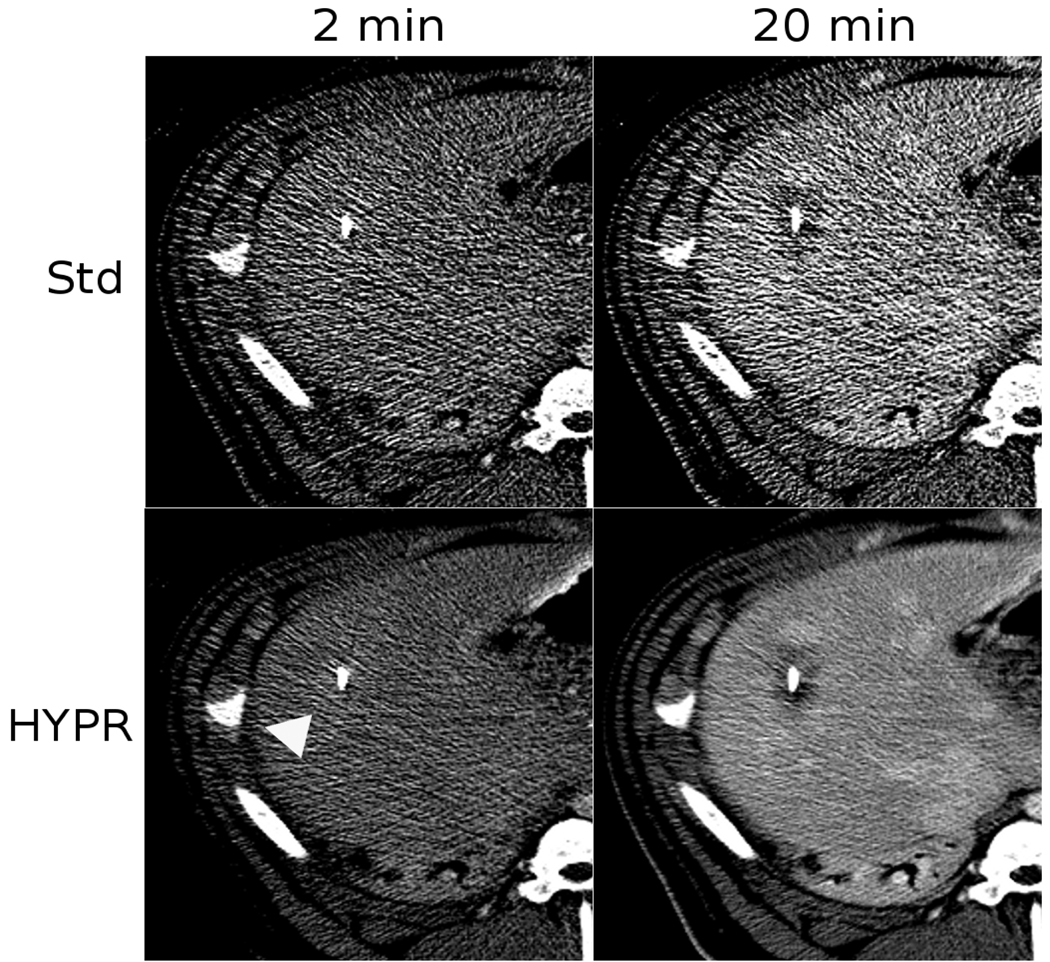
Figure 2.
Image comparison of standard reconstruction versus HYPR post-processing after 2 and 12 min of heating. In this case, a slight contrast-enhancement was observed after 2 min of heating due to local blood flow increases (arrow). As the zone of coagulation grew, enhancement was replaced by a darker area of low-attenuation. Note that the HYPR-processed images contain less noise and more clearly define the ablation zone, which is darker than the enhancing liver. Blood vessels are clearly visible in the 20 min HYPR image but not in the standard image.

