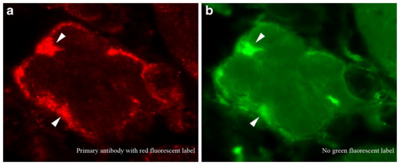
Fig. 1.

The potential for misinterpretation of artifact as protein aggregates in inclusion body myositis (IBM) myofibers. Immunofluorescence microscopy using a primary antibody (not disclosed here) with a red fluorochrome (a). The IBM literature is filled with similar images interpreted as demonstrating the accumulation of a specific protein. The same image viewed through a green fluorescent filter set (b), however, confirms that the material producing most, if not all, red fluorescence seen in (a) also emits green fluorescence. Because no green fluorescent molecules were used in this experiment, this material is autofluorescent. No protein aggregates can be concluded to be present. Had these studies been performed with a second green fluorescent-labeled antibody, they may have been interpreted incorrectly as showing colocalization of two different proteins in IBM aggregates. Arrowheads highlight some of the autofluorescent regions
