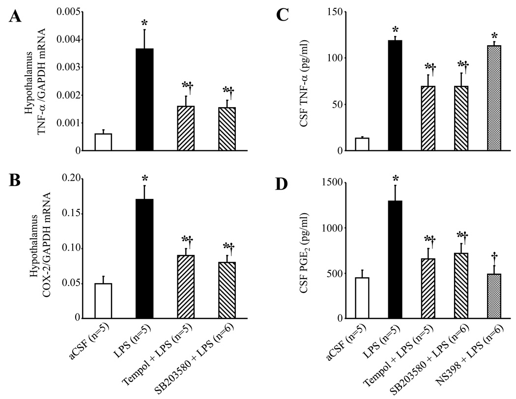
Fig. 4.
ICV administration of LPS increased hypothalamic tissue TNF-α (A) and COX-2 (B) mRNA expression, and both were significantly reduced by continuous ICV infusion of tempol and SB203580. ICV LPS also significantly increased cerebrospinal fluid (CSF) level of TNF-α (C) and PGE2 (D). ICV infusion of tempol and SB203580 reduced the LPS-induced increases in CSF TNF-α and PGE2; the COX-2 inhibitor NS398 reduced the LPS-induced increase in CSF PGE2. *P<0.05 versus ICV aCSF, †P<0.05, ICV Treatment + ICV LPS versus ICV LPS alone.

