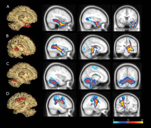
Figure 2.

Probabilistic DTI tractography showed abnormal white matter clusters found in TBSS linked to multiple distinct white matter tracts. DTI tractography was performed on each abnormal white matter cluster found in TBSS (shown in 3D glass brain, left side of the figure). Blue color indicates regions of the tract in which few subjects have in common, while red/yellow colors shows regions in which most subjects have in common. The anterior temporal cluster (A) was part of the uncinate fasciculus and inferior longitudinal fasciculus. The mesial temporal cluster (B) was associated with fornix, inferior longitudinal fasciculus, and motor projection tracts. The cerebellum cluster (C) had projections to bilateral anterior cerebellum. Tracking of the frontal parietal cluster (D) showed arcuate fasciculus and motor projection tracts.
