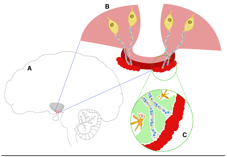
Figure 1.
Schematic representation of the hypothalamic-pituitary axis components that are involved in the control of reproduction. Panel A: The locations of the base of the hypothalamus (pink) and median eminence (dark red) are shown in reference to the brain as a whole. At the next layer of magnification (panel B), four GnRH cell bodies are depicted in yellow, with their nerve terminals projecting through the median eminence (dark red) to the portal capillaries (light red). Panel C is the highest magnification of the GnRH axons terminating at the portal capillaries. Tanycytes (green) are interspersed between GnRH and other terminals, and microglia (dark yellow) are shown. The blue circles in the axons and terminals in panels B and C represent large and small secretory vesicles. As the organization of the median eminence in this schematic is linear and the portal capillary vasculature basal lamina border is regular, this drawing depicts the situation in young rats. In aged rats (not shown) these relationships become disorganized.

