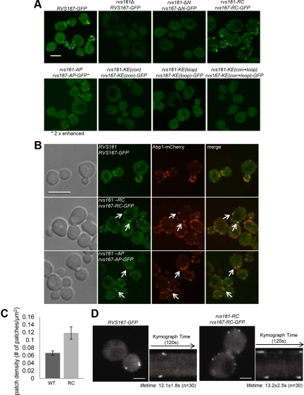
Figure 5.
Localization of Rvs167-GFP in rvs161 rvs167 mutants. (A) Maximum intensity projections of Z-stacks of C-terminally GFP-tagged Rvs167 strains. The signal for Rvs167-AP-GFP is two-fold enhanced to show its cortical localization. Scale bar, 4 μm. (B) Single images of Rvs167-GFP, Rvs167-RC-GFP, and Rvs167-AP-GFP with actin patch marker, Abp1-mCherry. Scale bar, 4 μm. (C) Rvs167-GFP patch density [patch number/cell surface area (μm2) ± SD] in wild-type and rvs161-RC rvs167-RC cells (n ≥ 100, three independent repeats). (D) Rvs167-RC-GFP exhibits a specific defect in its internalization step. On the left, single frames from live-cell movies show localization of Rvs167-GFP and Rvs167-RC-GFP. The corresponding kymographs are shown on the right. Movies corresponding to these kymographs are in Supplementary Movie 1 and 2. Each kymograph shows the dynamics of two or four patches on opposite sides of a cell over 120 s. Scale bar, 2 μm.

