Figure 7.
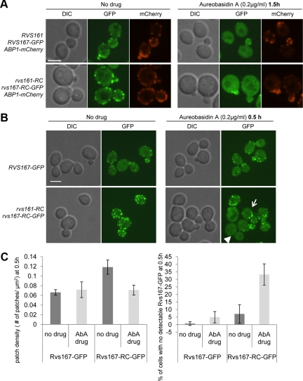
Rvs167-GFP localization is perturbed by aureobasidin A (AbA), an inhibitor of complex sphingolipid synthesis. (A) Localization of Rvs167-GFP and Abp1-mCherry in the absence (left) or in the presence of 0.2 μg/ml AbA was examined after 1.5 h of incubation by confocal microscopy in wild-type and RC-RC mutant cells. Single images were taken; scale bar, 4 μm. (B) RVS167-GFP and rvs161-RC rvs167-RC-GFP cells were treated with 0.2 μg/ml AbA for 0.5 h and imaged. Images show maximum intensity projections of multiple Z-series. Arrow indicates an example of a cell with a low number of GFP patches under AbA treatment. Arrowhead points to a cell where almost all patches have mislocalized. Scale bar, 4 μm. (C) Left, Rvs167-GFP patch density [number of patches/cell surface area (μm2)] in RVS167-GFP and rvs161-RC rvs167-RC-GFP cells after 0.5-h treatment with ± 0.2 μg/ml AbA. Right, quantification of Rvs167-GFP mislocalization in the presence of AbA show percentage of cells with no detectable Rvs167-GFP in the presence or absence of 0.2 μg/ml AbA at 0.5 h. n ≥ 100 cells, repeated three times.

