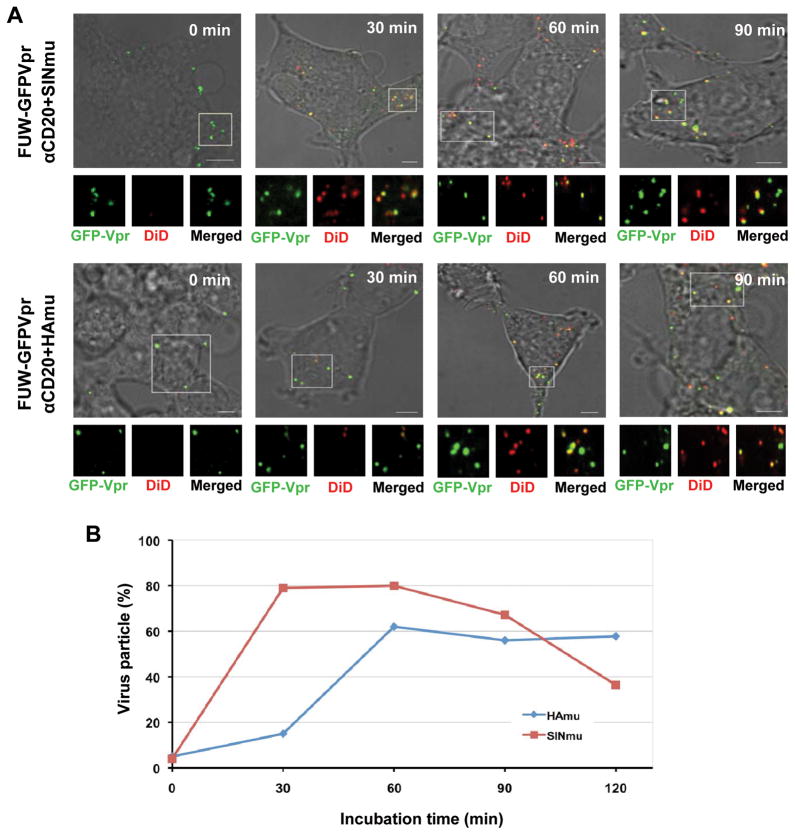
Fig. 3.
Visualization of virus-endosome fusion at different time points for engineered lentiviruses displaying SINmu or HAmu. (A) GFP-Vpr-labeled engineered lentiviruses (FUW-GFPVpr/αCD20+SINmu or FUW-GFPVpr/αCD20+HAmu; green) were labeled with DiD (red) for 1 h at room temperature. Double-labeled viruses were added to 293T/CD20 cells at 4°C for 30 min to synchronize the binding. The cells were then warmed to 37°C for 0, 30, 60, or 90 min, then fixed and imaged. The boxed regions are magnified and shown below in individual panels. Yellow signals indicate viral particles fused with endosomes. Scale bar represents 5 μm. (B) Quantification of the fused viral particles at different incubation time periods. GFP-Vpr+ viral particles with the fusion signal were quantified by viewing more than 100 viral particles at each time point. The viral particles positive for both GFP-Vpr and DiD were considered to be fused with endosomes, whereas particles that were only GFP-Vpr+ were considered to be unfused viruses. The results were collected from three independent experiments.

