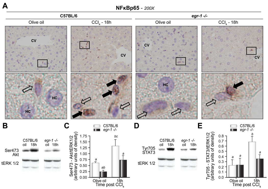
Fig. 5. Nuclear localization of NFκB-p65 in NPC-HS, and phosphorylation of Akt and STAT3 were reduced in egr-1−/− mice after CCl4 exposure.
(A) Immunohistochemistry was utilized to localize p65 in liver sections from wild-type and egr-1−/− mice. Closed arrows indicate p65-negative nuclei, while open arrows indicate p65-positive nuclei. CV = central vein; HC = hepatocyte. Outlined areas in (A) are shown enlarged below each 200X image. Images are representative of n = 4 – 8 per experimental group. Immunoblots of total hepatic protein were performed to determine expression of (B,C) phospho (Ser473)-Akt, (D,E) phospho (Tyr705)-STAT3 in livers from wild-type and egr-1−/− mice. Total (t)-Erk1/2 was used as a loading control. Representative (B) Ser473-Akt and (D) Tyr705-STAT3 immunoblots are shown. Quantification of band intensity measured by densitometry after normalization to tErk1/2 are shown in the bar graphs for (C) Ser-473 Akt and (E) Tyr705-STAT3 from n = 4–5 mice per group. Bars are means +/− SEM determined after scanning densitometry of immunoreactive bands.

