
Figure 4.
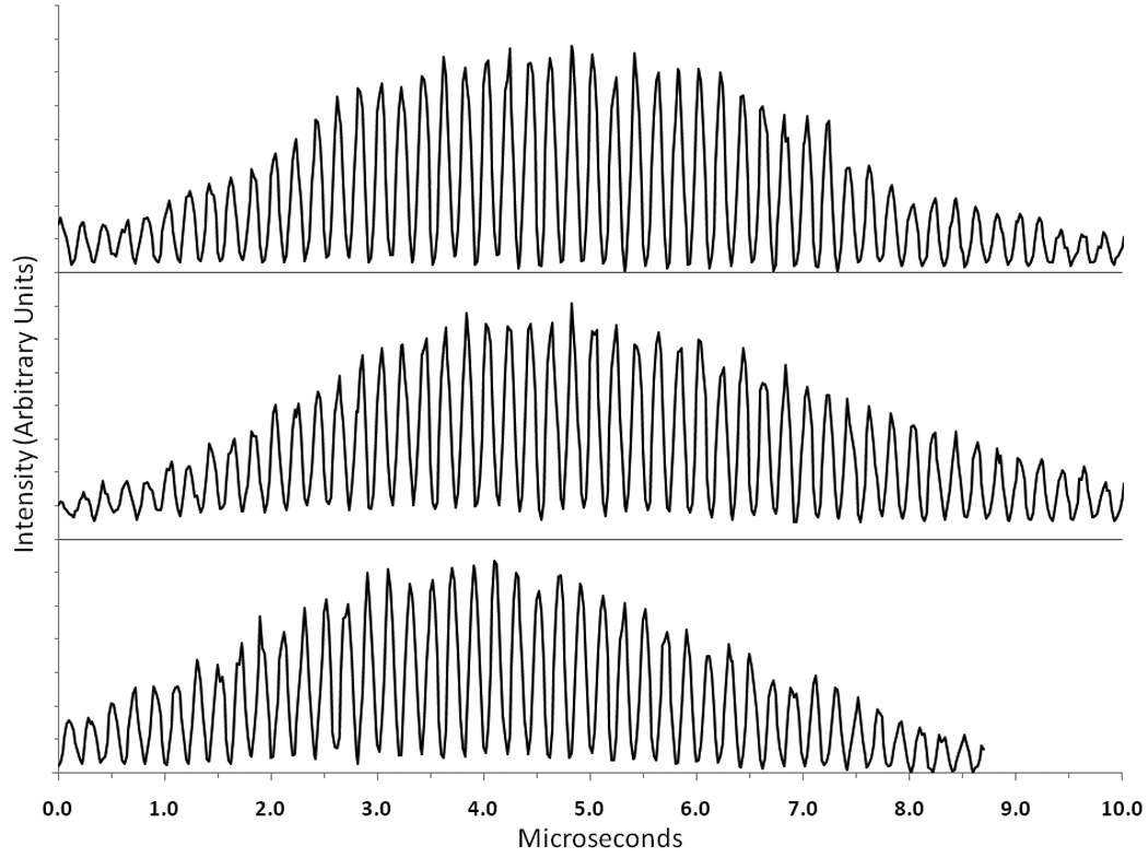
Examples of modulated waveforms detected for single particles using the digital system (y-axis is in arbitrary intensity units and individual traces are offset for clarity). The top trace is the 90° scatter signal for a single non-fluorescent bead, the middle trace is the fluorescence signal for a single Flow-Check™ bead and the bottom trace is the fluorescence signal for a single CHO cell stained with ethidium bromide.