
Fig. 6.
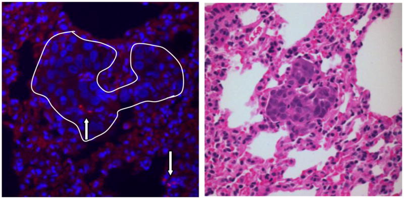
Specific localization of hUCMSCs in H358 tumor-bearing mouse lung. One week after three weekly injections of SP-DiI labeled hUCMSCs (5 × 10⁵ cells) to H358 tumor-bearing mice, mice were sacrificed and lungs were subjected to histochemical analysis. The fluorescent micrograph shows nuclei (blue) stained by Hoechst 33342 and selective engraftment of SP-DiI labeled hUCMS cells (A, red fluorescent cells indicated by arrows) in the H358 lung nodule (white line). Panel B shows an H and E stained serial section of tumor nodule.