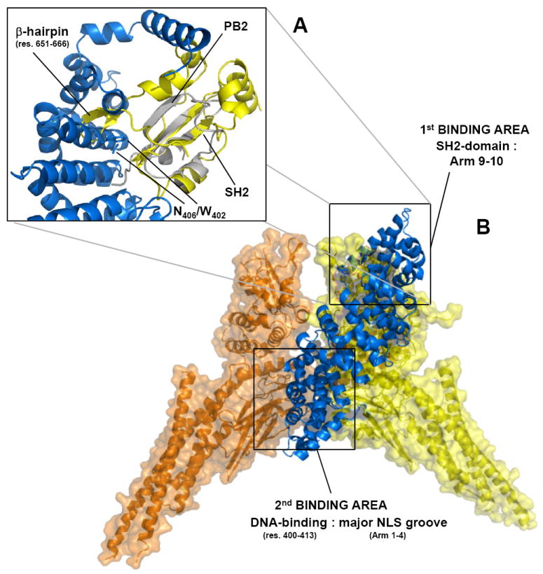
Figure 6. A structural model for the pSTAT1:importin α5 nuclear import complex.

(A) Superimposition of influenza PB2 subunit bound to importin α5 (in gray and blue, respectively) with STAT1–SH2 domain (in yellow). The rmsd deviation (on the α-carbon) between the PB2 globular domain (res. 686-757) and pSTAT1 SH2-domain (res. 558-634) is ~2.3 Å. (B) Structural model of dimeric pSTAT1 bound to one equivalent of importin α5. Importin α5 is shown as ribbon and colored in blue. Dimeric pSTAT1 is also in depicted in ribbon that is overlaid to a semi-transparent surface colored in orange and yellow for the two identical pSTAT1 protomers.
