FIGURE 3.
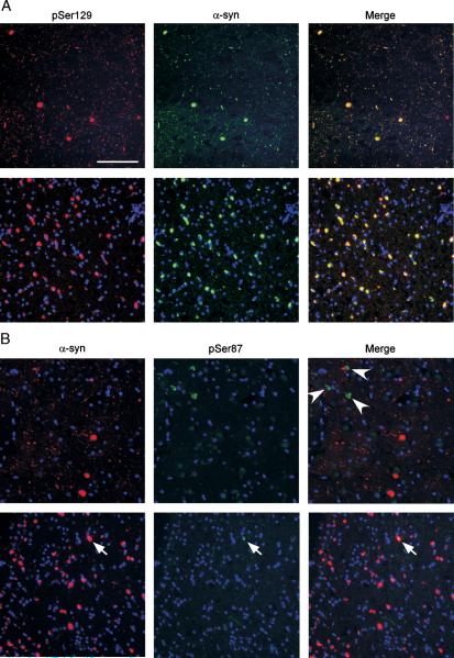
Double immunofluorescence with α-synuclein (α-syn) and phospho-specific antibodies in brain sections. (A) Tissue sections from the cingulate cortex from a patient diagnosed with Lewy body (LB) variant of Alzheimer disease (LBVAD; top row) or the cerebellum from a patient with multiple systems atrophy (MSA; bottom row) were double labeled with pSer129 (red) and α-syn (SNL-4 antibody; green). Extensive colocalization in LBs, Lewy neurites, and glial cytoplasmic inclusions (GCIs) was observed. (B) Tissue sections from the cingulate cortex from a patient with LBVAD (top row) or the cerebellum from a patient with MSA (bottom row) were double labeled with α-syn (Syn514; red) and pSer87 (green). Most LBs, Lewy neurites, and GCIs labeled with Syn514 were not immunoreactive with pSer87. Only occasional (<1%) GCIs labeled with Syn514 were also labeled with pSer87 (arrow). However, pSer87 labeled neurofibrillary tangle-like inclusions in cases of LBVAD (arrowheads). Scale bar = 100 μm.

