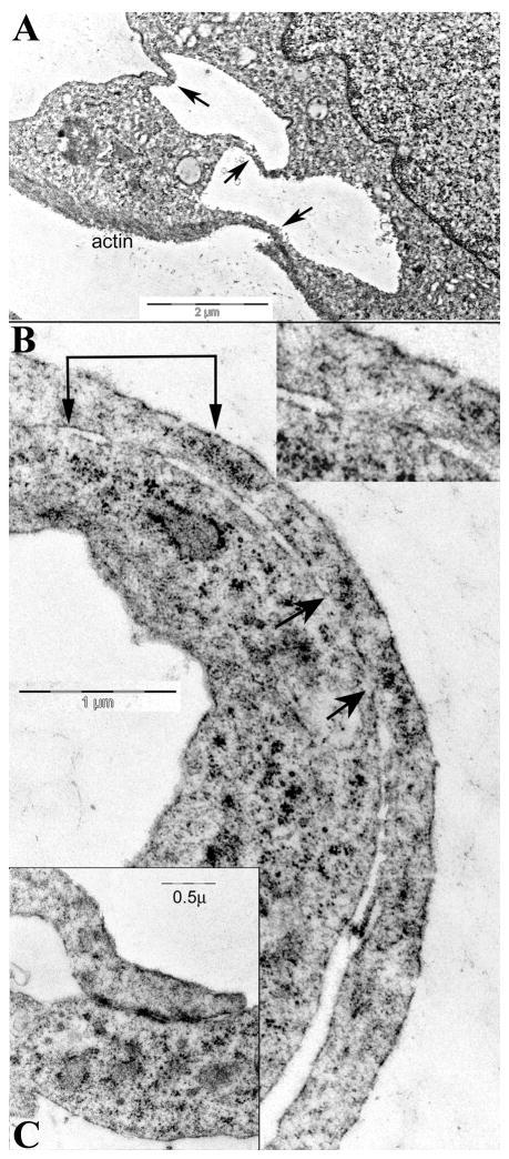
Fig. 4.
Representative ultrastructural images of contacts and processes in arrested cells. Fig. 4A shows three thin processes connecting two cells (arrows), and one has actin at its bottom surface. In B, two regions appear to share cytoplasm (at 2 connected arrows, magnified 2x in inset). Another larger region with cytoplasmic connection is also between the unconnected arrows. In C, a characteristic nanotube forms an adherent junction with another cell process that contains small vesicles.

