
Figure 1.
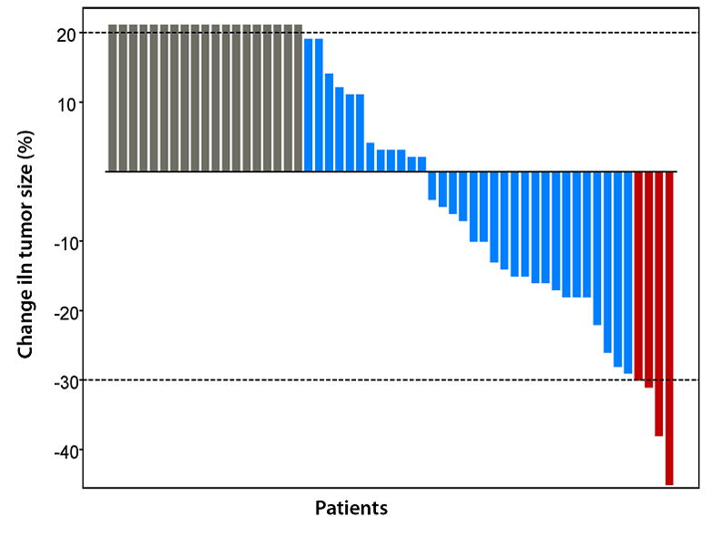
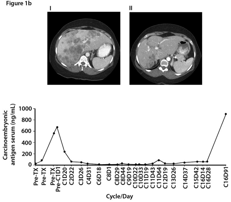
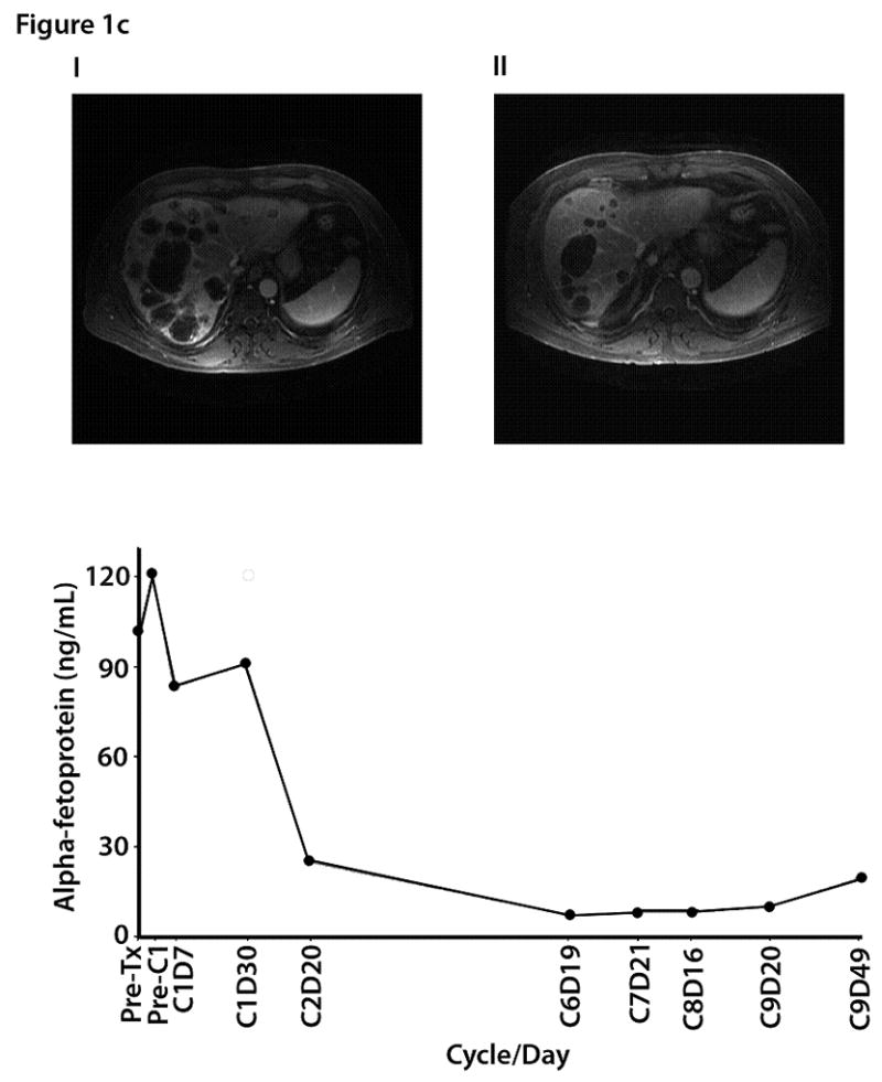
Figure 1a. Waterfall plot showing best response by RECIST to HAI oxaliplatin and systemic 5-FU, leucovorin, and bevacizumab: changes from baseline in tumor measurements. Each red box indicates a patient with maximum response, or a PR (> 30% reduction in tumor size) (n=4), each blue box indicates patients with SD (maximum response between 29% reduction and 19% increase in tumor size) (n = 32), and each grey box indicates a patient with clinically PD, or an increase in tumor > 20% (n=19).
Figure 1b. Computed tomography imaging from a 43-year-old female with colorectal carcinoma and liver metastases: (i) baseline (10/27/06) images demonstrate bilobar liver metastases and periportal adenopathy; (ii) after 8 months of treatment (06/07/07), improvement in the size and number of diffuse hepatic metastases was noted; (iii) carcinoembryonic antigen serum (CEA) levels by time of treatment.
Figure 1c. Magnetic resonance imaging (MRI) from a 51-year-old male with metastatic hepatocellular carcinoma: (i) baseline images demonstrated metastatic disease involving both lobes of the liver, right anterior diaphragmatic lymphadenopathy, and lower paraesophageal lymphadenopathy; (ii) 6 months later the size and number of multiple liver metastases had decreased; (iii) alpha fetoprotein trend levels by time of treatment.