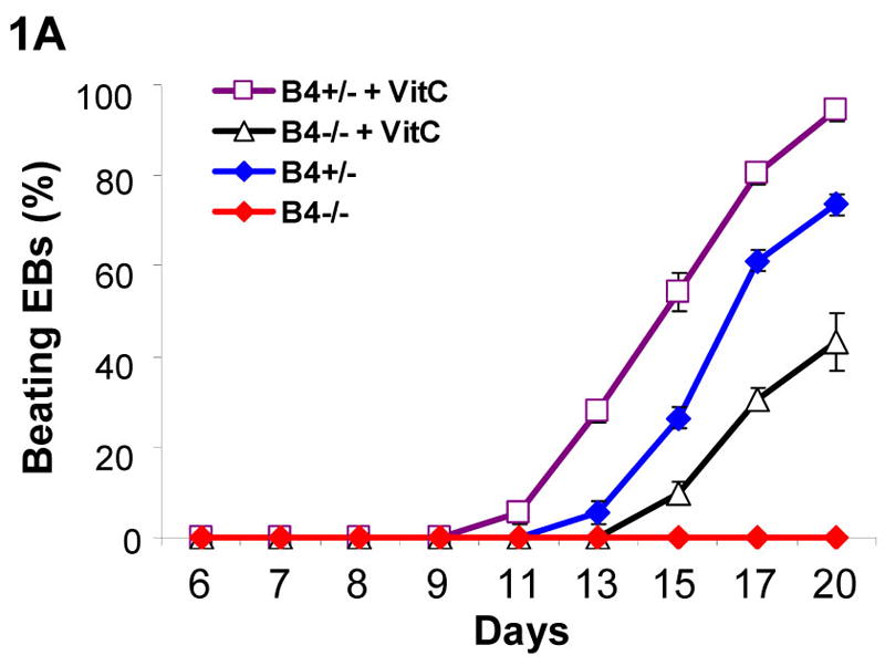
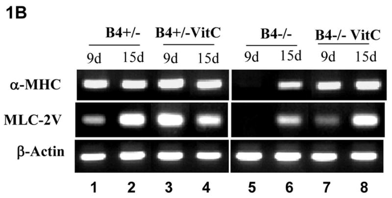
Figure 1. Defect of cardiomyocyte differentiation in EphB4-null ES cells.


Four hundreds of undifferentiated ES cells were differentiated in hanging-drops. Individual EBs were transferred to gelatin-coated wells at day 5. Spontaneous contracting EBs (beating EBs) were observed after EB attachment. (A) Undifferentiated EphB4+/− ES cells (B4+/−) or EphB4−/− ES cells (B4−/−) formed EBs in hanging-drop with or without ascorbic acid (VitC). After 5 days, individual EBs were transferred to 48-well plates. Beating EBs were counted at different time points. For each example, percentile of beating EB was calculated from total of 24 wells. Data are representative from 3 independent experiments. (B) RT-PCR analysis of cardiac genes, α-MHC and MLC-2V, in EphB4+/− or EphB4−/− ES cells. ES cells were differentiated in the presence or absence of ascorbic acid for 5 days. RNA samples were harvested at day 9 or day 15.
