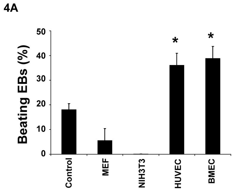
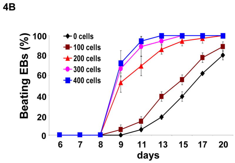
Figure 4. Effects of endothelial cells on cardiomyocyte differentiation.


(A) Undifferentiated α-MHC-GFP ES cells formed EBs in hanging drops (400 ES cells/drop) without 200 exogenous cells (control) or with MEF, NIH3T3, HUVEC, or BMEC cells. After 5 days, individual EBs were transferred to individual wells in 24 wells coated with gelatin. The wells containing beating EBs were counted at day 13 of differentiation. (B) Undifferentiated ES cells formed EBs in hanging-drops (400 ES cells/drop) containing 0, 100, 200, 300, or 400 BMEC cells. The wells containing beating EBs were counted on different days. Data are representative from 3 independent experiments. Error bars represent standard deviation (* indicates p<0.05 vs. control).
