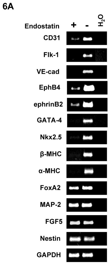
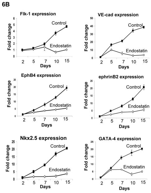
Figure 6. Expression of endothelial and cardiomyocyte genes during ES cell differentiation.

Differentiation of αMHC-GFP ES cells was carried out without (control) or with 2 μg/ml endostatin. (A) At day 10 of ES cell differentiation, RNA samples were harvested from EBs in the presence (+) or absence (−) of endostatin. RT-PCR was used to analyze gene expression. (B) RNA samples were harvested from EBs in the presence (Endostatin) or absence (Control) of endostatin at day 2, 5, 7, 10, and 15 of differentiation. Real-time PCR was performed with the primers listed in supplemental table 2. GAPDH was used as a standard.

