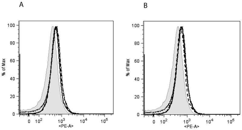
Figure 2. TLR4 expression in CD11b+ splenocytes (A) and bone marrow cells (B) from wild type, β-arrestin-1 and −2 KO mice.

Cell surface expression of TLR4 in CD11b+ splenocytes and bone marrow cells was determined by Flow-cytometry as described in the Methods. A representative experiment (showing the mean fluorescence intensity) from two such experiments is shown. Wild type=dotted line; β-arrestin-1 KO=dashed line and β-arrestin-2 KO=solid line; Isotype control (wild type)=shaded.
