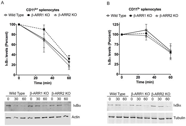
Figure 8. Regulation of IκBα levels by β-arrestins in CD11b+ (A) and CD11b− (B) splenocytes.
Splenocytes were obtained as described in the methods. Cells were then treated with LPS for the indicated time points and IκBα levels determined by Western blotting as described in the methods. IκBα levels were normalized to tubulin/actin for quantitation. N=4 mice per genotype. Quantitation is shown in the top and representative blots in the bottom.

