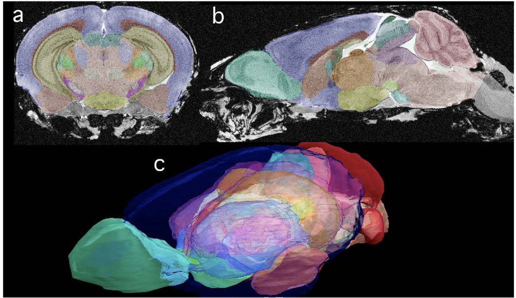
Figure 3.
All the MR data are isotropic. Thus, there is no loss of resolution between (a) the coronal plane and (b) the sagittal plane of CT2*. The color labels superimposed in (a) and (b) can be seen as transparent surfaces demonstrating the 3D juxtaposition of structure in the volume rendered image in (c). The key for the labels is included in Table 2.

