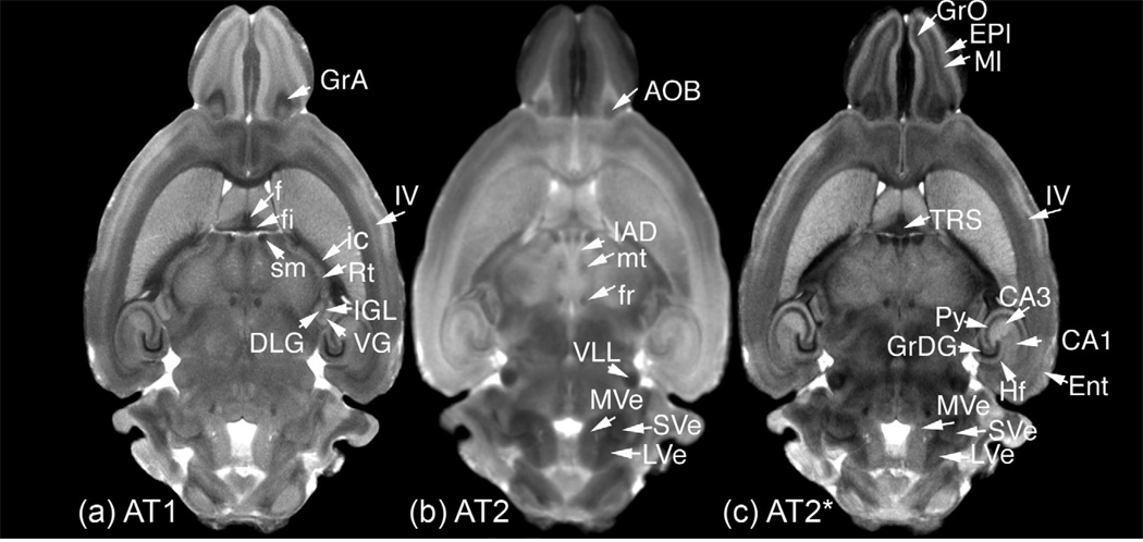
Figure 5.
The high contrast-to-noise-ratio provided in the average images: (a) AT1, (b) AT2, and (c) AT2* allow identification of a number of structures. Granule cell layers stand out in AT1 and AT2* as dark regions in the olfactory bulb (GrO - granular olfactory bulb, GrA - granular accessory olfactory), and hippocampus (GrDG - granular dentate gyrus). In contrast to GrDG, the pyramidal cell layer (Py) of the hippocampus appears less hypointense. A distinction between the CA1 (hypointense) and CA3 (hyperintense) areas of the hippocampus is most evident in the AT2*, but can also be seen in the AT1. White matter fibers such as fimbria (fi), fornix (f), fasciculus retroflexus (fr), internal capsule (ic), and stria medullaris (sm) are identified easily in the AT1. The mammillary tract is visible in the AT2. Thalamic (IAD - interanterodorsal, Rt - reticular, SG - suprageniculate, VG - ventral geniculate, DLG - dorsal lateral and IGL intrageniculate lamina) and hindbrain (MVE - medial-vestibular and LVE - lateral-vestibular) nuclei are best seen in AT2, as well as the nuclei of the lateral lemniscus (VLL - ventral nuclei of the lateral lemniscus) the accessory olfactory bulb (AOB). TRS - triangular septal nucleus, however, is best seen in the T2s. Differences in cell types, density, and myelination identify distinct areas of the cortex such as ENT- the entorhinal cortex, and individual layers, such as EPl - the external plexiform layer, MI - mitral cell layers in the olfactory areas, or Layer IV in the neocortex.

