
Fig 4.
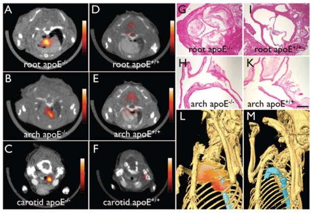
Non-invasive in vivo PET imaging of macrophages with a 64Cu-labeled trireporter nanoparticle [64Cu]-TNP. [64Cu]-TNP facilitates PET-CT imaging of inflammatory atherosclerosis in apoE−/− mice. Fused PET-CT images of the aortic root (A), arch (B), and carotid artery (C) of aged apoE−/− mice show strong PET signal in these vascular territories with high plaque burden, whereas no activity is observed in the same vasculature of wildtype mice (D through F). G and H, Hematoxylin and eosin histology of respective vascular regions, which carry a high plaque burden in apoE−/− but not in wild-type mice (I through K) (magnification: x 400 for G and I, x200 for H and K; bar=0.4 mm). The 3-dimensional maximum intensity reconstruction of the fused data set (L) demonstrates focal PET signal (red) in the proximal thoracic aorta (blue) of an apoE−/− mouse but not in a wild-type mouse (M). Reproduced from [88] with pending permission from the American Heart Association.