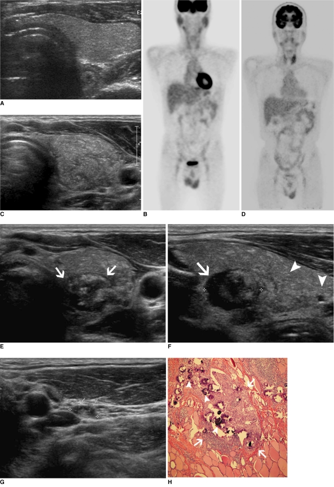
Fig. 1.
Diffuse sclerosing variant of papillary thyroid carcinoma in 48-year-old man.
A, B. Initial thyroid ultrasonography (A) and PET scan (B) have normal appearance.
C, D. Second-round screening examination was performed two years later. Ultrasonography (C) shows diffuse enlargement of left thyroid gland with heterogeneous echogenicity and formation of suspicious mass. It is regarded as pseudo-mass by heterogeneous parenchyma of left thyroid. PET scan (D) shows increased FDG uptake in both thyroid glands, especially in left lobe. These findings are regarded as benign thyroid disease and call for recommended follow-up examination.
E, F. Follow-up examination was performed six months later. Ultrasonography shows well defined cystic and solid mass (arrows) measuring 16 mm at left thyroid gland (E, F). It also shows multiple internal microcalcifications within this mass and multiple high echoic dots suggesting microcalcifications (arrowheads in F).
G. Lymph node of left level III shows nodular cortical thickening and microcalcifications. Metastasis was confirmed by surgery.
H. Photomicrograph shows mass (arrows) with multiple internal psammoma bodies (arrowheads) (Hematoxylin and Eosin staining, ×20).

