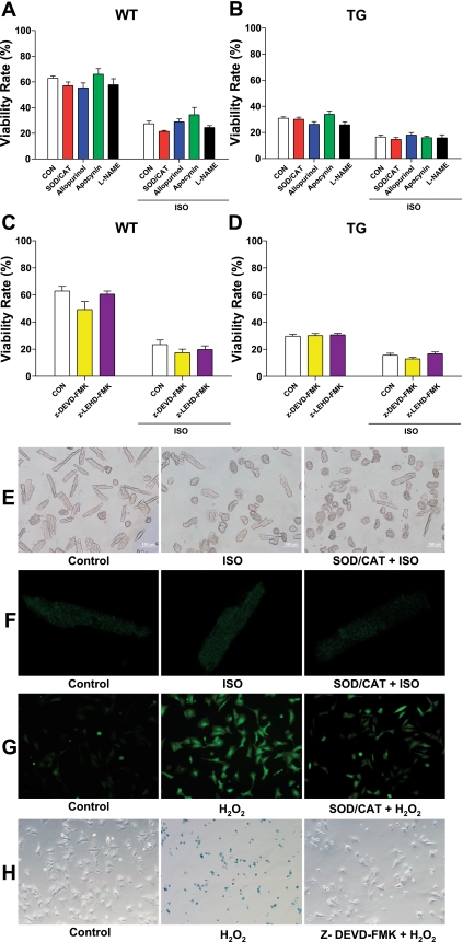
Fig. 5.
Inhibition of reactive oxygen species (ROS), nitric oxide synthase (NOS) pathways, and the caspase cascade did not affect ISO-induced WT and TG myocyte death. A and B: WT and TG myocyte viability, respectively, after 12 h of ISO treatment in the presence of ROS scavengers superoxide dismutase (SOD)-polyethylene glycerol (PEG) plus catalase (CAT)-PEG, ROS inhibitor allopurinol and apocynin, and NOS inhibitor NG-nitro-l-arginine methyl ester (l-NAME). C and D: WT and TG myocyte viability, respectively after 12 h of ISO treatment in the presence of caspase-3 inhibitor z-DEVD-fmk and caspase-9 inhibitor z-LEHD-FMK. Values are means ± SE. E: representative images of myocyte viability in WT adult mouse cardiomyocytes at 12 h. F: representative images of ROS production in WT adult mouse cardiomyocytes at 12 h, indicated by relative fluorescence intensity of carboxy-H2DCFDA. ISO and SOD-CAT treatment did not significantly alter the ROS production in rod-shaped myocytes. G: representative images of ROS production in neonatal rat ventricular myocytes (NRVMs). SOD-CAT reduced ROS production stimulated with H2O2 by 45.5 ± 6.2%. H: representative images of Trypan blue staining in NRVMs. Caspase-3 inhibitor z-DEVD-fmk reduced NRVM death induced with H2O2 by 55.7 ± 2.4%. N = 3.

