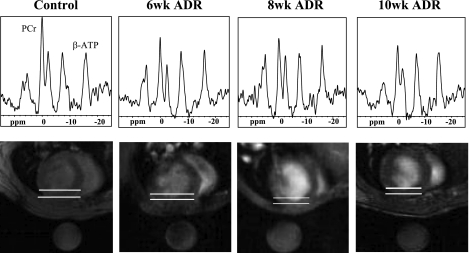
Fig. 3.
31P spectra (top) from the anterior myocardium, as indicated by the region between the white lines on the images (bottom), are shown with the prominent peaks of phosphocreatine (PCr) and β-phosphate of ATP (β-ATP). ppm, Parts/million. The round object below the animal in each image is a fiducial 31P standard contained within the probe. The cardiac PCr-to-ATP ratio (PCr/ATP) declines at 6 wk of ADR administration.

