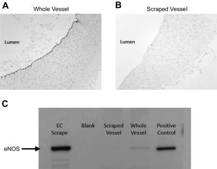
Fig. 1.
A: photomicrograph showing immunoreactive staining for endothelial nitric oxide synthase (eNOS) along the endothelial lining of an intact femoral artery. B: photomicrograph demonstrating loss of immunoreactivity to eNOS after scraping the luminal surface of the artery to remove endothelium. C: Western blot demonstrating enrichment of endothelial protein content with mechanical scraping of the luminal surface (femoral artery). Note the increase in eNOS immunoreactivity between scraped vessel, whole vessel, and endothelial scrape. EC, endothelial cell.

