Fig. 2.
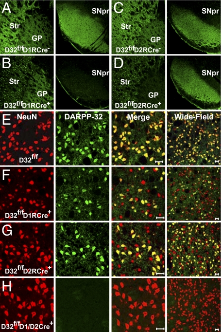
Selective deletion of DARPP-32 in striatonigral or striatopallidal neurons. (A–D) Immunofluoresence performed on brain sections from D32f/fD1RCre− (A), D32f/fD1RCre+(B), D32f/fD2RCre− (C), and D32f/fD2RCre+ (D) mice using an antibody against DARPP-32. (Left) Somatic DARPP-32 staining in the striatum (Str) and axonal labeling in the GP. (Right) Corresponding axonal DARPP-32 labeling in the SNpr. (E–H) Immunofluoresence of striatal sections from D32f/f (E), D32f/fD1RCre+ (F), D32f/fD2RCre+ (G), and D32f/fD1/D2RCre+ mice (H) costained with antibodies against NeuN (red) and DARPP-32 (green). The third column shows a merged image, and the fourth column shows a merged image of a different brain section at lower magnification. (Scale bar: 20 μm.)

