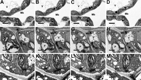
Fig. 3.
Focused ion beam (FIB) electron micrographs of 13-d-old leaf tissue of WT (Top row), PEX10-ΔZn1 (Middle row) and PEX10-G93E (Bottom row). The tissue was stained for catalase activity with diaminobenzidine. Images at successive levels of sections exposed by the ion beam are shown. (A–D) Leaf peroxisomes of WT plants are ovoid and in physical contact with chloroplasts. (E–H) Leaf peroxisomes of PEX10-ΔZn1 are worm-like and accumulate at places distant from chloroplasts. (I–M) Leaf peroxisomes of PEX10-G93E plants are elongated but are attached to chloroplasts. Mb, microbody (peroxisome); P, chloroplasts; V, vacuole. (Scale bar, 3 μm.)

